Background
 Pelvic
inflammatory disease (PID) is an infectious and inflammatory disorder
of the upper female reproductive tract, including the uterus, fallopian
tubes, and adjacent pelvic structures.
Pelvic
inflammatory disease (PID) is an infectious and inflammatory disorder
of the upper female reproductive tract, including the uterus, fallopian
tubes, and adjacent pelvic structures. Pelvic inflammatory disease (PID) is initiated by infection that ascends from the vagina and cervix. Chlamydia trachomatis is the predominant sexually transmitted organism causing PID. Newer, more accurate, laparoscopic studies have shown that PID may often be polymicrobial in nature (30-40%). Other organisms that have been implicated in the pathogenesis of PID include Neisseria gonorrhoeae, Gardnerella vaginalis, Haemophilus influenzae, and anaerobes, such as Peptococcus and Bacteroides species. (see Etiology).
At presentation, women with PID may range from asymptomatic to seriously ill. The most common presenting complaint is lower abdominal pain. Many women also exhibit an abnormal vaginal discharge. The diagnosis of acute PID is primarily based on historical and clinical findings, but many patients may exhibit only a few or no symptoms. (See Clinical Presentation.)
The classic high-risk patient is a menstruating woman younger than 25 years who has multiple sex partners, does not use contraception, and lives in an area with a high prevalence of sexually transmitted disease (STD).
The differential diagnosis includes appendicitis, cervicitis, urinary tract infection, endometriosis, and adnexal tumors. PID is the most common incorrect diagnosis in cases of ectopic pregnancy. A pregnancy test is required in all women of childbearing age. A delay in diagnosis or treatment of PID can result in long-term sequelae, such as chronic pelvic pain and tubal infertility. (See Differentials.)
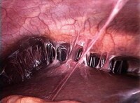
PID may produce tubo-ovarian abscess (TOA) and extend to produce pelvic peritonitis and Fitz-Hugh-Curtis syndrome (perihepatitis), as shown in the image below.
"Violin-string" adhesions of chronic Fitz-Hugh-Curtis syndrome. Laparoscopy is the current criterion standard for the diagnosis of PID. No single test is highly specific or sensitive for the disease, but certain laboratory studies that can be used to support the diagnosis include the erythrocyte sedimentation rate, C-reactive protein, and chlamydial and gonococcal DNA probes and cultures. Imaging studies, such as ultrasound, computed tomography, and magnetic resonance imaging may also prove helpful in unclear cases. (See Workup.)
Empirical treatment is suggested by the Centers for Disease Control and Prevention (CDC) Sexually Transmitted Disease Management Guidelines in patients with uterine or adnexal tenderness and cervical motion tenderness, if no other etiology explains the findings. All antibiotic regimens must be effective against C trachomatis and N gonorrhoeae, as well as against gram-negative facultative organisms, anaerobes, and streptococci . Most patients are now treated in an outpatient setting, but physicians should consider hospitalization in selected cases. (See Treatment and Management.)
Anatomy
Pelvic
inflammatory disease may extend from infection of the lower female
reproductive tract, including the vagina and cervix. Pelvic inflammatory
disease (PID) is an infectious and inflammatory disorder of the upper
female reproductive tract, including the uterus and fallopian tubes.
Infection and inflammation may spread to adjacent pelvic structures in
the pelvis and abdomen, including perihepatic structures (Fitz-Hugh
Curtis syndrome).
Pathophysiology
Most
cases of pelvic inflammatory disease (PID) are presumed to occur in 2
stages. The first stage is acquisition of a vaginal or cervical
infection; this infection is often sexually transmitted and may be
asymptomatic. The second stage is direct ascent of microorganisms from
the vagina or cervix to the upper genital tract, with infection and
inflammation of these structures.
The exact mechanism of ascent of microorganisms from the vagina and cervix is unknown. However, studies have suggested that a number of factors may be involved. Although cervical mucus provides a functional barrier against upward spread, the efficacy of this mechanism may be decreased by hormonal changes that occur during ovulation and menstruation.
Alterations in the cervicovaginal microenvironment may also result from antibiotic treatment and sexually transmitted infections that can disrupt the balance of endogenous flora, causing normally nonpathogenic organisms to overgrow and ascend. Opening of the cervix during menstruation with retrograde menstrual flow may also facilitate ascent of microorganisms.
Intercourse may contribute to the ascent of infection due to rhythmic mechanical uterine contractions. Bacteria may be carried along with sperm into the uterus and tubes
It has also been suggested that genetic polymorphisms of PID pathogens affect the likelihood that a lower tract infection will progress to frank PID. Chlamydial heat shock protein 60 (CHSP60) antigen expression in C trachomatis[1] and P9Opa(b) protein expression in N gonorrhoeae[2] are examples of specific bacterial genes implicated in the pathology of PID.
In the upper tract, a number of microbial and host factors appear to play a role in the degree of host inflammation and resultant scarring. Tubal infection initially affects the mucosa, but acute, complement-mediated transmural inflammation may develop rapidly and increase in intensity with subsequent infections.
Inflammation may extend to uninfected parametrial structures, including the bowel. Infection may extend by spillage of purulent materials from the fallopian tubes or via lymphatic spread beyond the pelvis to produce acute peritonitis and acute perihepatitis (Fitz-Hugh Curtis syndrome).
Pregnancy-related factors
Pregnancy decreases the risk of PID once the cervical os is protected by the mucous plug. PID rarely occurs in pregnancy; however, the disease can occur in the first 12 weeks of gestation, before the mucous plug solidifies and seals off the uterus from ascending bacteria; fetal loss may result. Concurrent pregnancy influences the choice of antibiotic therapy for PID and demands that an alternative diagnosis of ectopic pregnancy be excluded. Uterine infection is usually limited to the endometrium but may be more invasive in a gravid or postpartum uterus.Genetics
Den Hartog et al found a possible contributing role of 5 single-nucleoside polymorphisms (SNPs) in 4 genes encoding pattern recognition receptors in local tubal cells and circulating immune cells (eg, macrophages). The presence of 2 or more SNPs in patients appeared to correlate with increased laparoscopically identifiable tubal pathology.[3]Etiology
Infecting organisms
The organisms most commonly isolated in many, if not most, cases of acute PID are Neisseria gonorrhoeae and Chlamydia trachomatis.[4] C trachomatis, an intracellular bacterial pathogen, is the predominant sexually transmitted organism causing PID, In the United States, the role of N gonorrhoeae as the primary cause of PID has decreased; however, it remains the second most frequently reported sexually transmitted infection after Chlamydia. C linically, infection may be asymptomatic or manifest similarly to Chlamydia. An estimated 10-20% of untreated chlamydial or gonorrheal infections progress to PID.However, newer studies using more sensitive and specific laparoscopic cultures have found acute PID to be polymicrobial in up to 30-40% of cases. N gonorrhoeae and C trachomatis may be instrumental in the initial infection of the upper tract, with anaerobes, facultative anaerobes, and other bacteria increasingly isolated as inflammation increases and abscesses form. Organisms involved include the following:
- Mycoplasma hominis
- Mycoplasma genitalium[5]
- Ureaplasma urealyticum
- Herpes simplex virus–2 (HSV-2)
- Trichomonas vaginalis
- Cytomegalovirus
- Haemophilus influenza
- Streptococcus agalactiae
- Enteric gram-negative rods (Escherichia coli)
- Peptococcus species
- Anaerobes
The microbiology of PID has also been found to reflect the predominant sexually transmitted infections (STIs) prevalent within a specific population and also less-common organisms seen in that population. Bacterial vaginosis (BV) is suggested to play a role in the initiation of ascending infection in a subset of women with heavy growth of BV-associated organisms, such as G vaginalis, more than 2 recent sexual partners, and especially after recent abortion or gynecologic surgery.[6, 7] In less-developed countries, PID may be due to a granulomatous salpingitis caused by Mycobacterium tuberculosis or Schistosoma species.[8]
Patients infected with T vaginalis demonstrated a 4-fold increase in the histologic evidence of acute endometritis in a 2006 cross-sectional study of 736 women with PID. Co-infection of HSV-2 with N gonorrhoeae, C trachomatis, and BV was also associated with histologic evidence of acute endometritis. HSV-2 was demonstrated to be associated with fallopian tube inflammation and lower tract ulcerations that may contribute to disruption of the endocervical canal mucus barrier.[9]
Human immunodeficiency virus (HIV) infection has been found to be associated with an increased incidence of C trachomatis infection, Candida, and human papillomavirus. Women with HIV infection also have an increased risk of progression to PID and tubo-ovarian abscess.[10]
Microbial virulence appears to play a significant role in PID. Bjartling et al studied different chlamydial strains recovered from patients with PID and found less symptomatic disease in infection produced by a less virulent variant strain.[11]
Risk factors
Risk factors for PID include multiple sexual partners, a history of prior STIs, and a history of sexual abuse.[12] Frequent vaginal douching has also been implicated. Frequent vaginal douching has been considered a risk factor for PID,[13] but studies reveal no clear association.[14]Younger age has been found to be associated with increased risk, suggested to be due to some combination of increased cervical mucosal permeability, a larger zone of cervical ectopy, a lower prevalence of protective chlamydial antibodies, and increased risk-taking behaviors. Surgical procedures, such as endometrial biopsy, curettage, and hysteroscopies break the cervical barrier, predisposing women to ascending infections.
The microbiology of PID has also been found to reflect the predominant STIs prevalent within a specific population and also less-common organisms seen in that population. Bacterial vaginosis (BV) is suggested to play a role in the initiation of ascending infection in a subset of women with heavy growth of BV-associated organisms, such as G vaginalis, more than 2 recent sexual partners, and especially after recent abortion or gynecologic surgery.[6, 7] PID may result from Mycobacterium tuberculosis in endemic areas.[8]
Contraception
Different forms of contraception may affect PID incidence and severity. Appropriately used barrier contraception has clearly been shown to decrease the acquisition of most STIs.The CDC recommends that spermicides and condoms containing nonoxynol-9 should be avoided, as a number of African studies have demonstrated that nonoxynol-9 can cause vaginal lesions and may increase the risk of HIV transmission. While the level of nonoxynol-9 in condoms is lower than the level associated with vaginal lesions, these are also not recommended because they are more expensive, have a shorter shelf life, and have been associated with urinary tract infections.[15]
Studies of oral contraceptive pills (OCPs) have found differing effects on PID risks. On the one hand, OCPs are thought to increase the risk of endocervical infection, probably by increasing the zone of cervical ectopy. On the other hand, evidence has indicated that OCPs can decrease the risk of symptomatic PID, possibly by increasing cervical mucus viscosity, decreasing menstrual anterograde and retrograde flow, and modifying local immune responses. Still other data have suggested that OCPs may not have any effect on PID incidence.[15]
Intrauterine-device (IUD) use has been associated with a 2- to 9-fold increased risk for PID, but data suggest that the risk with current IUDs may be significantly less.[16] Kelly et al found a rate of 9.6 cases of PID per 1,000 IUD insertions, with the most significant risk in the first 20 days.[17] Meirik et al validated early risk of PID within the first month after insertion and also found that the risk appears to be modified by the patient’s number of sexual partners, patient’s age, and the community prevalence of STIs.[18]
Actinomycete species have been identified almost exclusively in patients with IUDs.[19]
Bilateral tubal ligation (BTL) has not been found to provide protection against PID; however, patients with BTL may have delayed or milder forms of PID.[20]
Epidemiology
From 1995-2001, 769,859 cases of PID were reported in the United States annually.[21] The
true incidence was probably much higher; cases likely went unreported
due to incomplete and untimely conventional, nonelectronic reporting
methods and because many cases of silent and smoldering PID occur and
are discovered only when the patient develops chronic complications.
The CDC has estimated that more than 1 million women experience an episode of PID every year. The disease leads to approximately 2.5 million office visits and 125,000-150,000 hospitalizations yearly.[22, 23]
Worldwide, WHO has determined that STIs rank in the top 5 disease categories for which adults seek care. Women in resource-poor countries, especially those in sub-Saharan Africa and Southeast Asia, experience an increased rate of complications and sequelae.
The annual rate of PID in high-GNP countries has been reported to be as high as 10-20 per 1000 women of reproductive age. Public health efforts implemented in Scandinavia to decrease the prevalence of STIs have been quite effective.
The CDC has estimated that more than 1 million women experience an episode of PID every year. The disease leads to approximately 2.5 million office visits and 125,000-150,000 hospitalizations yearly.[22, 23]
International statistics
While no specific international data are available for PID incidence worldwide, the World Health Organization (WHO) estimated in 1999 that approximately 340 million new cases of curable STIs occur annually in individuals aged 15-49 years.[24] Factors contributing to the difficulty in determining the actual worldwide incidence and prevalence of PID include lack of patient recognition of disease, difficulties in access to care, the often subjective method of disease diagnosis, lack of diagnostics and laboratory facilities in many developing countries, and underfunded and overstretched public health systems.[25]Worldwide, WHO has determined that STIs rank in the top 5 disease categories for which adults seek care. Women in resource-poor countries, especially those in sub-Saharan Africa and Southeast Asia, experience an increased rate of complications and sequelae.
The annual rate of PID in high-GNP countries has been reported to be as high as 10-20 per 1000 women of reproductive age. Public health efforts implemented in Scandinavia to decrease the prevalence of STIs have been quite effective.
Prognosis
Chronic
pelvic pain occurs in approximately 25% of patients with a history of
pelvic inflammatory disease (PID). This pain is thought to be related to
cyclic menstrual changes, but it also may be the result of adhesions or
hydrosalpinx. Impaired fertility is a major concern in women with a history of PID. Infection and inflammation can lead to scarring and adhesions within tubal lumens. Of women with tubal factor infertility (TFI), 50% have no history of PID but have scarring of the fallopian tubes and exhibit antibodies to C trachomatis. The rate of infertility increases with the number of episodes of infection. The risk of ectopic pregnancy is increased 15-50% in women with a history of PID. Ectopic pregnancy is a direct result of damage to the fallopian tube.
PID may produce tubo-ovarian abscess (TOA) and extend to produce pelvic peritonitis and Fitz-Hugh Curtis syndrome (perihepatitis), as shown in the image below. TOA is reported in up to one third of women hospitalized for PID.
"Violin-string" adhesions of chronic Fitz-Hugh-Curtis syndrome. Approximately 125,000-150,000 hospitalizations occur yearly because of PID.[22] Women in resource-poor countries, especially those in sub-Saharan Africa and Southeast Asia, experience an increased rate of complications and sequelae.
Patient Education
Asking
women about high-risk sexual behavior is very important. Encourage
screening tests for those at risk. Additionally, ensure that male sex
partners are evaluated and treated.
Patient education should focus on methods of preventing PID and STIs, including reducing the number of sexual partners, avoiding unsafe sexual practices, and routinely using appropriate barrier protection. Adolescents should be advised to delay the onset of sexual activity until age 16 years or older, as they are at an increased risk for PID.
After treatment, women should be counseled to abstain from sexual activity or be educated to strictly and appropriately use barrier protection until their symptoms have fully abated and they have completed their antibiotic regimen and their partner(s) have been treated.
For patient education information, see the Women's Health Center, Sexually Transmitted Diseases Center, and Pregnancy and Reproduction Center, as well as Pelvic Inflammatory Disease, Ectopic Pregnancy, Birth Control Overview, Birth Control FAQs, and Female Sexual Problems.
Rebound lower abdominal tenderness and involuntary guarding may be noted and suggest associated peritonitis. The positive predictive value (PPV) of these findings will vary depending on the prevalence of PID in a given population.
One large, multicenter trial found adnexal tenderness to be the most sensitive physical examination finding (95% sensitive; P < .001).[28] Mucopurulent cervicitis is common, and if absent, it provides a significant negative predictive value (NPV). Adnexal fullness or disproportionate unilateral adnexal tenderness may indicate the development of a tubo-ovarian abscess.
Molander et al found the following 3 variables to be significant predictors of the diagnosis, correctly classifying 65% of patients with laparoscopically documented PID (95% confidence interval, 61-99%)[29] :
Patient education should focus on methods of preventing PID and STIs, including reducing the number of sexual partners, avoiding unsafe sexual practices, and routinely using appropriate barrier protection. Adolescents should be advised to delay the onset of sexual activity until age 16 years or older, as they are at an increased risk for PID.
After treatment, women should be counseled to abstain from sexual activity or be educated to strictly and appropriately use barrier protection until their symptoms have fully abated and they have completed their antibiotic regimen and their partner(s) have been treated.
For patient education information, see the Women's Health Center, Sexually Transmitted Diseases Center, and Pregnancy and Reproduction Center, as well as Pelvic Inflammatory Disease, Ectopic Pregnancy, Birth Control Overview, Birth Control FAQs, and Female Sexual Problems.
History
The
classic high-risk patient is a menstruating woman younger than 25 years
who has multiple sex partners, does not use contraception, and lives in
an area with a high prevalence of sexually transmitted infections
(STIs).
Pelvic inflammatory disease (PID) is more prevalent among individuals who are young at first intercourse. Additionally, the IUD confers a relative risk of 2.0-3.0 for the first 4 months following insertion, but then it decreases to baseline thereafter. Women who are not sexually active have a very low incidence of upper genital tract infection, as do women who have undergone total abdominal hysterectomy. Bilateral tubal ligation (BTL) does not provide protection against PID; however, patients post BTL may have delayed and milder forms of the disease.
Depending on the severity of the infection, patients with PID may be minimally symptomatic or may present with toxic symptoms of fever, nausea, vomiting, and severe pelvic and abdominal pain.
Gonococcal PID is thought to have an abrupt onset with more toxic symptoms than nongonococcal disease. Gonorrhea- and chlamydia-associated infections are more likely to cause symptoms toward the end of menses and in the first 10 days following menstruation.
Lower abdominal pain is present. Usually, pain is described as dull, aching or crampy, bilateral, and constant; it begins a few days after the onset of the last menstrual period and tends to be accentuated by motion, exercise, or coitus. Pain from PID usually lasts less than 7 days; if pain lasts longer than 3 weeks, the likelihood that PID is the correct diagnosis declines substantially.
Abnormal vaginal discharge is present in approximately 75% of cases, and unanticipated vaginal bleeding, often postcoital, coexists in about 40% of cases.[26]
Temperature higher than 38°C (30% of cases), nausea, and vomiting manifest late in the clinical course of the disease.
Physical Examination
Because of the serious potential complications of untreated PID and the endemic prevalence of the infection, the Centers for Disease Control and Prevention (CDC) has adopted an approach to maximize diagnosis by using minimal criteria and by urging providers to maintain a low threshold for diagnosis and empiric treatment. Institute empiric treatment of PID when a sexually active young woman who is at risk for STI has pelvic or lower abdominal pain, no identifiable cause for her illness other than PID, and, on pelvic examination, 1 or more of the following minimal criteria[27] :- Cervical motion tenderness
- Uterine tenderness
- Adnexal tenderness
Rebound lower abdominal tenderness and involuntary guarding may be noted and suggest associated peritonitis. The positive predictive value (PPV) of these findings will vary depending on the prevalence of PID in a given population.
One large, multicenter trial found adnexal tenderness to be the most sensitive physical examination finding (95% sensitive; P < .001).[28] Mucopurulent cervicitis is common, and if absent, it provides a significant negative predictive value (NPV). Adnexal fullness or disproportionate unilateral adnexal tenderness may indicate the development of a tubo-ovarian abscess.
Molander et al found the following 3 variables to be significant predictors of the diagnosis, correctly classifying 65% of patients with laparoscopically documented PID (95% confidence interval, 61-99%)[29] :
- Adnexal tenderness (P < .001)
- Fever (P < .001)
- Elevated sedimentation rate (ESR) (P < .001)
Diagnostic Considerations
The diagnosis of acute pelvic inflammatory disease (PID) is primarily based on historical and clinical findings. The diagnostic process is imprecise, with no single piece of historical, physical, or laboratory information found to be highly specific or sensitive for the disease.Patients may be asymptomatic with endocervical infections and PID. Uncomplicated endocervical infections with C trachomatis and N gonorrhoeae are underdiagnosed and tend to be undertreated.[31] Bjartling et al have found less symptomatic urethral infection and decreased lower abdominal findings produced by a less virulent variant strain of C trachomatis.[11]
Although many patients with PID have atypical presentations and exhibit no or few symptoms, more than 25% of these patients meet objective criteria for upper tract infection on laparoscopic examination. The sensitivity of the pelvic examination is only 60%.
Due to the relatively poor specificity and sensitivity of clinical findings, the CDC has established minimal criteria for the diagnosis of PID. Institute empiric treatment of PID when a patient who is at risk for sexually transmitted disease (STD) has pelvic or lower abdominal pain, no identifiable cause for her illness other than PID, and, on pelvic examination, 1 or more of the following minimal criteria[27] :
- Cervical motion tenderness
- Uterine tenderness
- Adnexal tenderness
All female patients of childbearing age with lower abdominal pain require a pregnancy test. PID is the most common incorrect diagnosis in missed ectopic pregnancy.
Pain from PID usually lasts less than 7 days; if pain lasts longer than 3 weeks, the likelihood that PID is the correct diagnosis declines substantially.
Most patients show clinical response within 48-72 hours after medical therapy. If the patient continues to have fever, chills, uterine tenderness, adnexal tenderness, and cervical motion tenderness, consider other possible causes and a diagnostic laparoscopy.
Differential Diagnoses
- Adnexal Tumors
- Appendicitis
- Ectopic Pregnancy
- Endometriosis
- Ovarian Cysts
Approach Considerations
A
number of procedures can be performed to improve the diagnosis of
pelvic inflammatory disease (PID) and its complications. These
procedures are not necessary, nor are they indicated, in the management
of every case of PID. However, due to the difficulty of definitive
clinical diagnosis and the number of important surgical and gynecologic
emergencies that may have similar presentations, the clinician should be
aware of these modalities. Specific criteria for PID based on
procedures that may be appropriate for some patients are as follows:
Additional criteria that improve diagnostic specificity include the following:
- Laparoscopic confirmation
- Transvaginal ultrasonographic scanning or magnetic resonance imaging (MRI) showing thickened, fluid-filled tubes with/without free pelvic fluid or tubo-ovarian abscess (TOA)
- Endometrial biopsy showing endometritis
Additional criteria that improve diagnostic specificity include the following:
- Oral temperature greater than 38.3° C (101° F)
- Abnormal cervical or vaginal mucopurulent discharge
- Abundant white blood cells (WBCs) on saline microscopy of vaginal secretions
- Elevated erythrocyte sedimentation rate
- Elevated C-reactive protein
- Laboratory evidence of cervical infection with N gonorrhoeae or C trachomatis (culture or DNA probe)
Lab Studies
Perform
a pregnancy test. If the results are positive, the possibility of
ectopic pregnancy must be addressed. This also directly influences
antibiotic choice and consideration of the patient for admission.
On a complete blood count (CBC), less than 50% of women with acute PID have a WBC count above 10,000. Due to the poor sensitivity and specificity, an elevated WBC count is not a CDC criterion for diagnosing PID.
In fact, no single test is highly specific and sensitive for PID; however, a number of tests may be used to increase the specificity of the clinical diagnosis. Saline and potassium hydroxide–treated preparations of vaginal secretions can be examined for leukorrhea (>10 WBC/high-power field, >1 WBC/epithelial cell), trichomoniasis, and clue cells.[5, 33] The presence of leukorrhea was found to be the most sensitive, but not specific, laboratory indicator of upper tract infection; the absence of leukorrhea is a negative predictor of PID.
Other nonspecific findings include elevation of the erythrocyte sedimentation rate (ESR), C-reactive protein (CRP), or WBC count.
Gonorrhea DNA probes and cultures are generally used to support the diagnosis and to provide epidemiologic data for public health departments, but they are frequently negative in later stages. Chlamydial DNA probes and cultures are generally used to support the diagnosis and to provide epidemiologic data for public health departments, although there is large variability in recovery from the cervix (5-56%). Quantitative culture for chlamydia identifies rapidly replicating bacteria that appear to be associated with active disease. However, DNA probe and culture results are often not available to the emergency physician at the time of initial evaluation.
One study suggested that women with a high titer of IgG chlamydial antibodies, acute pelvic pain, and a clinical picture suggestive of PID were more likely to have salpingitis than adhesions alone. Those patients with high titers and chronic pelvic pain, but with a clinical picture that did not suggest PID, were more likely to have adhesions alone. The investigators concluded that their limited data suggested that serologic testing might help to formulate the diagnosis.[34]
Other tests that may be considered include the following:
- Rapid protein reagin (RPR) test for syphilis (syphilis is again increasing in the United States)
- Hepatitis and HIV
- Urinalysis to help exclude urinary tract infections (however, a positive urinalysis does not exclude PID, because any inflammatory process in the contiguous pelvis can produce white blood cells in the urine)
Transvaginal Ultrasonographic Scanning
Ultrasonographic
scanning is one diagnostic imaging examination performed in cases of
suspected PID in which clinical findings are nondiagnostic. Transvaginal
ultrasonography is superior to transabdominal ultrasonography for
diagnosing PID, as well as endometrial abnormalities and pelvic masses.[33] This
modality is readily available and noninvasive and can be performed at
the patient's bedside. There are no large randomized trials addressing
the specificity and sensitivity of bedside ultrasonography in PID
diagnosis. The literature demonstrates that the sensitivity and
specificity depend on the criteria used to indicate PID, the quality of
the equipment, and the experience of the individual operator performing
the test.
Transvaginal ultrasonography has poor sensitivity (81%) and specificity (78%) in mild or atypical PID.[33] Helpful findings include thickened (>5 mm), fluid-filled fallopian tubes; indistinct endometrial borders; ovaries with multiple small cysts; and moderate-to-large amounts of free pelvic fluid in acute, severe PID. Small amounts of free pelvic fluid have not been shown to be a discriminatory finding. These findings alone do not demonstrate adequate specificity to make a definitive diagnosis of PID.
In the patient who appears toxic or has asymmetric pelvic findings, ultrasonographic scanning is an important diagnostic tool for the identification of a TOA. Pelvic abscesses may be seen as complex, adnexal masses with multiple internal echoes. The modality has been shown to demonstrate as many as 70% of adnexal masses missed on physical examination.
Pelvic ultrasonographic scanning (see the images below) is also useful in evaluating the possibility of ectopic pregnancy in patients whose differential diagnosis includes that condition and PID. The modality can also be helpful in evaluating other disorders in the differential diagnosis, including hemorrhagic ovarian cyst, ovarian torsion, endometrioma, and appendicitis. The use of ultrasound appears to be medical center specific, as some adult academic medical centers do not believe that ultrasound is of appropriate sensitivity and specificity to be used as a solo imaging modality to rule out appendicitis.
 Transabdominal
ultrasonogram. This image shows anechoic tubular structures in the
adnexa; the finding is compatible with a hydrosalpinx.
Transabdominal
ultrasonogram. This image shows anechoic tubular structures in the
adnexa; the finding is compatible with a hydrosalpinx. 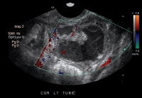 Endovaginal
ultrasonogram. This image reveals a tubular structure with debris in
the left adnexa; the finding is compatible with a pyosalpinx.
Endovaginal
ultrasonogram. This image reveals a tubular structure with debris in
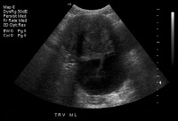
the left adnexa; the finding is compatible with a pyosalpinx. 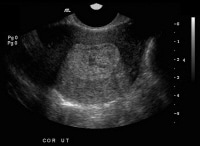 This ultrasonogram shows a markedly heterogeneous and thickened endometrium, a finding that is compatible with endometritis.
 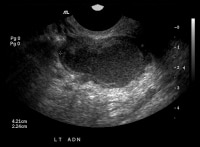 This
ultrasonogram reveals bilateral complex masses in a patient who had
pyometrium, a finding that is compatible with tubo-ovarian abscess.
This
ultrasonogram reveals bilateral complex masses in a patient who had
pyometrium, a finding that is compatible with tubo-ovarian abscess. 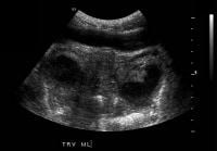 Transabdominal
ultrasonogram. This image demonstrates an echogenic region within the
endometrium with dirty shadowing, a finding that is compatible with air
in the endometrium and endometritis. Additionally, bilateral complex
masses are present; this finding is compatible with tubo-ovarian masses.
Ultrasonographic results in patients with PID may be normal
or nonspecific, because salpingitis alone is not usually associated with
imaging findings.[35]
Transabdominal
ultrasonogram. This image demonstrates an echogenic region within the
endometrium with dirty shadowing, a finding that is compatible with air
in the endometrium and endometritis. Additionally, bilateral complex
masses are present; this finding is compatible with tubo-ovarian masses.
Ultrasonographic results in patients with PID may be normal
or nonspecific, because salpingitis alone is not usually associated with
imaging findings.[35]
Positive ultrasonographic findings in PID may include the following:
Hydrosalpinx and pyosalpinx can usually be readily distinguished from pelvic veins and bowel by visualizing the color flow within the patent blood vessels and peristalsis within the bowel.
Imaging findings in TOAs are usually nonspecific and must be distinguished from endometriomas, ectopic pregnancies, hemorrhagic cysts, ovarian tumors, and abscesses from adjacent organs.
Transvaginal ultrasonography has poor sensitivity (81%) and specificity (78%) in mild or atypical PID.[33] Helpful findings include thickened (>5 mm), fluid-filled fallopian tubes; indistinct endometrial borders; ovaries with multiple small cysts; and moderate-to-large amounts of free pelvic fluid in acute, severe PID. Small amounts of free pelvic fluid have not been shown to be a discriminatory finding. These findings alone do not demonstrate adequate specificity to make a definitive diagnosis of PID.
In the patient who appears toxic or has asymmetric pelvic findings, ultrasonographic scanning is an important diagnostic tool for the identification of a TOA. Pelvic abscesses may be seen as complex, adnexal masses with multiple internal echoes. The modality has been shown to demonstrate as many as 70% of adnexal masses missed on physical examination.
Pelvic ultrasonographic scanning (see the images below) is also useful in evaluating the possibility of ectopic pregnancy in patients whose differential diagnosis includes that condition and PID. The modality can also be helpful in evaluating other disorders in the differential diagnosis, including hemorrhagic ovarian cyst, ovarian torsion, endometrioma, and appendicitis. The use of ultrasound appears to be medical center specific, as some adult academic medical centers do not believe that ultrasound is of appropriate sensitivity and specificity to be used as a solo imaging modality to rule out appendicitis.
Transabdominal
ultrasonogram. This image shows anechoic tubular structures in the
adnexa; the finding is compatible with a hydrosalpinx. Endovaginal
ultrasonogram. This image reveals a tubular structure with debris in
the left adnexa; the finding is compatible with a pyosalpinx. This ultrasonogram shows a markedly heterogeneous and thickened endometrium, a finding that is compatible with endometritis. This
ultrasonogram reveals bilateral complex masses in a patient who had
pyometrium, a finding that is compatible with tubo-ovarian abscess. Transabdominal
ultrasonogram. This image demonstrates an echogenic region within the
endometrium with dirty shadowing, a finding that is compatible with air
in the endometrium and endometritis. Additionally, bilateral complex
masses are present; this finding is compatible with tubo-ovarian masses.
Ultrasonographic results in patients with PID may be normal
or nonspecific, because salpingitis alone is not usually associated with
imaging findings.[35] Positive ultrasonographic findings in PID may include the following:
- The uterus may be ill defined because of inflammation; however, inflammation of the uterus is an unusual finding
- Endometritis may result in central-endometrial-cavity echo thickening and heterogeneity
- Hydrosalpinx is depicted as a fluid-filled fallopian tube (if the fallopian tube walls are thickened and if debris is present within the tube, pyosalpinx should be considered in the differential diagnosis, but a pyosalpinx may be imaged as an echoless tube, whereas an imaged echo-filled tube may be due to proteinaceous but noninfected fluid in a hydrosalpinx)
- Oophoritis results in enlarged ovaries with ill-defined margins that often appear adherent to the uterus; adjacent free fluid may be present in the adnexa or cul-de-sac
- Tubo-ovarian abscesses (TOAs) are depicted as complex adnexal masses with thickened walls and central fluid
- Pelvic infection, such as tubal hyperemia, detected by Doppler studies, is one of the most specific criteria in diagnosing PID.[36]
Hydrosalpinx and pyosalpinx can usually be readily distinguished from pelvic veins and bowel by visualizing the color flow within the patent blood vessels and peristalsis within the bowel.
Imaging findings in TOAs are usually nonspecific and must be distinguished from endometriomas, ectopic pregnancies, hemorrhagic cysts, ovarian tumors, and abscesses from adjacent organs.
Laparoscopy
Laparoscopy
is the criterion standard for the diagnosis of PID. It is significantly
more specific and sensitive than are clinical criteria alone. The
minimum criteria to diagnose PID laparoscopically include tubal wall
edema, visible hyperemia of the tubal surface, and the presence of
exudate on the tubal surfaces and fimbriae.
Pelvic masses consistent with tubo-ovarian abscess or ectopic pregnancy can be directly visualized. Hepatic abscess exudate and/or adhesions may be visible. Material can be obtained for definitive culture and histologic studies.
Drawbacks of laparoscopy are that the procedure is expensive and invasive, exhibits interobserver variability, and requires an operating room and anesthesia.[29] Findings on laparoscopy do not necessarily correlate with the severity of illness, as only the surfaces of structures are visible. Laparoscopy may not fully define PID in up to 20% of cases.
Computed Tomography
Computed
tomography (CT) scanning may also be used as the initial diagnostic
study for the investigation of nonspecific pelvic pain in a female, and
PID may be found incidentally. Ultrasonographic imaging is preferred
over CT scanning as the triaging tool in a female child or adolescent
with right lower quadrant or pelvic pain, because of concerns about
radiation exposure.
CT scan findings are nonspecific in cases of PID in which there is no evidence of an abscess. Inflammation obliterates the pelvic fat planes, with thickening of the fascial planes. If hydrosalpinx is present, a fluid-filled tubular structure may be seen in the adnexa.
Typically, a TOA is visualized as a mass; the mass may have regular margins and contain debris similar to that seen in endometriomas or hemorrhagic cysts. The margins may be thick and irregular. There may also be an associated low-attenuation area that may represent an adjacent or contained fluid-filled fallopian tube.[37] Many adult centers also prefer this modality to ultrasonography when a diagnosis of appendicitis is in question.
Tubular, fluid-filled, nonvascular structures in the pelvis that are associated with an adnexal mass are suggestive of dilated fallopian tubes that correlate with cases of PID. A finding of an adjacent or surrounding complex mass confirms the diagnosis of TOA.
Magnetic Resonance Imaging
Although the specificity (95%) and sensitivity (95%) of magnetic resonance imaging (MRI) are relatively high,[33] the modality is costly and rarely indicated in acute PID.
Hydrosalpinx is depicted as a tubular structure with low signal intensity on T1-weighted MRI scans and high signal intensity on T2-weighted images. If the walls are thickened, pyosalpinx should be considered in the differential diagnosis.[38]
Oophoritis may be evidenced by enlarged, polycystic-appearing ovaries with ill-defined margins and adjacent fluid.
TOAs often appear as thick-walled masses with low signal intensity on T1-weighted images and high signal intensity on T2-weighted images. Occasionally, the TOA may be isointense or hyperintense on T1-weighted images, and they may have heterogeneous signal intensity on T2-weighted images.
Culdocentesis
Culdocentesis
can be performed rapidly in the emergency department. With the advent
of transvaginal ultrasonographic scanning, culdocentesis is rarely
performed, but it is valuable in settings where current technology is
unavailable. For the procedure, an 18-gauge spinal needle attached to a
20-mL syringe is inserted transvaginally into the cul-de-sac. Normally,
this yields only 2-4 mL of clear to straw-colored free pelvic fluid;
purulent fluid indicates an infectious or inflammatory process. The
potential positive findings of leukocytes and bacteria are nonspecific
and may indicate PID or may be a product of another infectious or
inflammatory process in the pelvis, such as appendicitis or
diverticulitis, or may be due to contamination with vaginal contents. A
yield of more than 2 mL of nonclotting blood is consistent with ectopic
pregnancy.
Endometrial Biopsy
Endometrial
biopsy can be used to determine the histopathologic diagnosis of
endometritis, a condition that is uniformly associated with salpingitis.
Endometrial biopsy is approximately 90% specific and sensitive. The
procedure is performed with an endometrial suction pipette/curette and
is well tolerated. Specimens for culture may also be obtained during the
procedure, but these are frequently contaminated with vaginal flora.
The 2010 update to the CDC sexually transmitted diseases treatment guideline recommends endometrial biopsy in women undergoing laparoscopy who have no visible signs of salpingitis, since endometritis can be the only sign of PID.[36]
Diagnostic use of endometrial biopsy in the emergency department is limited due to the requirement for operator training. In addition, results are not immediately available to the clinician.
Improved education, routine screening, diagnosis, and empirical treatment of these infections should decrease the incidence and prevalence of these processes and the incidence of long-term sequelae. Education should concentrate on strategies to prevent PID and STIs, including reducing the number of sexual partners, avoiding unsafe sexual practices, and routinely using appropriate barrier protection. Adolescents should be advised to delay the onset of sexual activity until age 16 years or older, as they are at an increased risk for PID.
Women with PID should be counseled to abstain from sexual activity, or be educated to strictly and appropriately use barrier protection, until their symptoms and those of their partner[36] have fully abated and they have completed their entire treatment regimen.
Based on published data, the US Preventive Services Task Force (USPSTF) recommends screening for chlamydia in all sexually active, nonpregnant women up to age 25 years and in nonpregnant women aged 25 years or older who are at increased risk (grade A recommendation), as well as in all pregnant women up to age 25 years and in pregnant women aged 25 years or older who are at increased risk (grade B recommendation). The USPSTF recommends against routine screening for women aged 25 years and older, whether or not they are pregnant, if they are not at increased risk (grade C recommendation).
The USPSTF does not provide recommendations for chlamydia screening in men, due to insufficient evidence regarding benefits and risks.[46] However, a 2008 demonstration project suggested that the combination of partner notification and the screening of men with a relatively high prevalence of chlamydia and a larger number of partners would be more cost-effective than expanding screening to low-risk women.[47]
Patients treated for STIs and PID may be noncompliant with medication regimen because of low medical literacy and may not understand their diagnosis. These individuals frequently do not follow up or notify partners. Patients should be fully educated about these issues, as well as about the advisability of testing and treatment for other STIs, including HIV, hepatitis, and syphilis. In particular, the 2010 CDC guidelines state that HIV testing should be offered to all women diagnosed with acute PID.
The 2010 update to the CDC sexually transmitted diseases treatment guideline recommends endometrial biopsy in women undergoing laparoscopy who have no visible signs of salpingitis, since endometritis can be the only sign of PID.[36]
Diagnostic use of endometrial biopsy in the emergency department is limited due to the requirement for operator training. In addition, results are not immediately available to the clinician.
Approach Considerations
The
treatment of pelvic inflammatory disease (PID) addresses the relief of
acute symptoms, eradication of current infection, and minimization of
the risk of long-term sequelae. These sequelae, including chronic pelvic
pain, ectopic pregnancy, tubal factor infertility (TFI), and
implantation failure with in vitro fertilization attempts, may occur in
up to 25% of patients.[39]
From a public health perspective, treatment is aimed at the expeditious eradication of infection in order to reduce the risk of transmission of infection to new partners and to identify and treat current and recent partners to further help prevent sexually transmitted infection (STI).
Early diagnosis and treatment appears to be critical in the preservation of fertility. Current guidelines suggest that empirical treatment should be initiated in at-risk women who exhibit lower abdominal pain, adnexal tenderness, and cervical motion tenderness. Due to diagnostic difficulties and the potential for serious sequelae, the Centers for Disease Control and Prevention (CDC) advises that physicians maintain a low threshold for aggressive patient treatment, with overtreatment preferred to no or delayed treatment.
Therapy using antibiotics alone is successful in 33-75% of cases. If surgical treatment is warranted, the current trend in therapy is conservation of reproductive potential with simple drainage, adhesiolysis, and copious irrigation or unilateral adnexectomy, if possible. Further surgical therapy is needed in 15-20% of cases so managed.
Outpatient Versus Inpatient Treatment
Most
patients with PID are managed as outpatients, but physicians should
consider hospitalization for patients with the following conditions,
although no clear data suggest that these patients benefit from
hospitalization:
Most patients show clinical response within 48-72 hours after medical therapy. If the patient continues to have fever, chills, uterine tenderness, adnexal tenderness, and cervical motion tenderness, consider other possible causes and a diagnostic laparoscopy.
Admission of persons infected with HIV and of adolescents should be reviewed on an individual basis. Admission decisions are based on the following factors:
- Uncertain diagnosis
- Pelvic abscess on ultrasonographic scanning
- Pregnancy
- Failure to respond to outpatient management
- Inability to tolerate outpatient oral antibiotic regimen
- Severe illness or nausea and vomiting precluding outpatient treatment
- Immunodeficiency (eg, patients with HIV infection who have a low CD4 count, or patients using immunosuppressive medications)
- Failure to improve clinically after 72 hours of outpatient therapy
Most patients show clinical response within 48-72 hours after medical therapy. If the patient continues to have fever, chills, uterine tenderness, adnexal tenderness, and cervical motion tenderness, consider other possible causes and a diagnostic laparoscopy.
Admission of persons infected with HIV and of adolescents should be reviewed on an individual basis. Admission decisions are based on the following factors:
- Diagnostic certainty
- Illness severity
- Likelihood of compliance with outpatient regimen
- Whether or not the patient is pregnant
- Coexisting immunosuppression or illness
- Major fertility issues
- Risk factors for significant anaerobic infection (eg, IUD use, recent pelvic procedure, presence of TOA)
- Obstetrician/gynecologist
- Surgeon (especially if appendicitis or another intra-abdominal process cannot be excluded)
- Infectious disease consultant (especially in patients who are HIV positive and may be on highly active antiretroviral treatment [HAART])
Antibiotic Regimens
Treatment
initiated in the emergency department, clinic, or office setting should
be expeditiously begun and should include empirical broad-spectrum
antibiotics to cover the full complement of common causes. All regimens
must be effective against Chlamydia trachomatis and Neisseria gonorrhoeae, as well as against gram-negative facultative organisms, anaerobes, and streptococci.
A number of studies (1992-2006) have demonstrated the effectiveness of a variety of parenteral and oral regimens in the elimination of acute symptoms and in microbiologic cure.[32] No differences in outcome were identified between inpatient and outpatient management in a large, randomized, multicenter, NIH-sponsored clinical study that effectively compared inpatient and outpatient oral and parenteral antibiotic regimens in the documented elimination of endometrial and tubal infection.[42]
Physicians should be aware of current guidelines and current national and local patterns of drug resistance in their patient populations to avoid inappropriate treatment.[43] If an IUD is present, it should be removed after the initiation of antibiotic treatment.
Patients on an intravenous PID regimen can be transitioned to oral antibiotics 24 hours after clinical improvement. These should be continued for a total of 14 days. Oral therapy usually involves doxycycline (Vibramycin); however, azithromycin (Zithromax, Zmax) can also be used.[44] In patients who have developed TOA, oral therapy should include clindamycin (Cleocin) or metronidazole (Flagyl). (See Medication.)
All patients should be reevaluated in 72 hours for evidence of clinical improvement and compliance with their antibiotic regimen. Multiple studies have shown poor compliance with doxycycline therapy, and approximately 20-25% of patients have never filled their prescriptions.
Laparoscopy and Laparotomy
Patients
who do not improve in 72 hours should be reevaluated for possible
laparoscopic or surgical intervention and for reconsideration of other
possible diagnoses. Laparoscopic pelvic lavage, abscess drainage, and
adhesion lysis may be necessary.
Most TOAs (60-80%) resolve with antibiotic administration. If patients do not respond appropriately, laparoscopy may be useful for identifying loculations of pus requiring drainage. An enlarging pelvic mass may indicate bleeding secondary to vessel erosion or a ruptured abscess. Unresolved abscesses may be drained percutaneously via posterior colpotomy, via CT or ultrasonographic guidance, laparoscopically, or by laparotomy.
The advantages of laparoscopy include direct visualization of the pelvis and more accurate bacteriologic diagnosis if cultures are obtained. However, laparoscopy is not always available in acute PID. In addition, this procedure is costly and requires general anesthesia. It should be used if the diagnosis is in doubt. However, if operative laparoscopy is used early in the course of the disease, copious irrigation and separation of thin adhesions by blunt dissection may prevent later sequelae.
Laparotomy is usually reserved for surgical emergencies, such as abscesses that have ruptured or that have not responded to medical management and laparoscopic drainage, and for patients who are not candidates for laparoscopic management. Treatment is guided by intraoperative findings and the patient's desire for fertility maintenance. Treatment may involve unilateral salpingo-oophorectomy or hysterectomy and bilateral salpingo-oophorectomy. Ideally, surgery is performed after the acute infection and inflammation have resolved. In patients with recurrent PID, dense pelvic adhesions may render surgery difficult.
Deterrence and Prevention
Randomized, controlled trials suggest that preventing chlamydial infection reduces the incidence of PID.[45] In addition, all sexual partners of women with PID should be treated empirically for C trachomatis and N gonorrhoeae if they have had sexual contact with the patient in the 60 days preceding the onset of her symptoms. Additionally, the 2010 CDC guidelines recommend that if a patient last had sexual intercourse more than 60 days before onset of symptoms or diagnosis, the most recent sex partner should be treated. Urethral gonococcal or chlamydial infection in the partner is highly likely and is frequently asymptomatic in men. Even in clinical settings where men do not receive treatment, arrangements for care or referral of male sex partners should be made. Regardless of whether a woman’s sex partners were treated, women diagnosed with chlamydial or gonococcal infection should follow up with repeat testing within 3-6 months, as these women have a high rate of reinfectionwithin6months of treatment.[36]Improved education, routine screening, diagnosis, and empirical treatment of these infections should decrease the incidence and prevalence of these processes and the incidence of long-term sequelae. Education should concentrate on strategies to prevent PID and STIs, including reducing the number of sexual partners, avoiding unsafe sexual practices, and routinely using appropriate barrier protection. Adolescents should be advised to delay the onset of sexual activity until age 16 years or older, as they are at an increased risk for PID.
Women with PID should be counseled to abstain from sexual activity, or be educated to strictly and appropriately use barrier protection, until their symptoms and those of their partner[36] have fully abated and they have completed their entire treatment regimen.
Based on published data, the US Preventive Services Task Force (USPSTF) recommends screening for chlamydia in all sexually active, nonpregnant women up to age 25 years and in nonpregnant women aged 25 years or older who are at increased risk (grade A recommendation), as well as in all pregnant women up to age 25 years and in pregnant women aged 25 years or older who are at increased risk (grade B recommendation). The USPSTF recommends against routine screening for women aged 25 years and older, whether or not they are pregnant, if they are not at increased risk (grade C recommendation).
The USPSTF does not provide recommendations for chlamydia screening in men, due to insufficient evidence regarding benefits and risks.[46] However, a 2008 demonstration project suggested that the combination of partner notification and the screening of men with a relatively high prevalence of chlamydia and a larger number of partners would be more cost-effective than expanding screening to low-risk women.[47]
Patients treated for STIs and PID may be noncompliant with medication regimen because of low medical literacy and may not understand their diagnosis. These individuals frequently do not follow up or notify partners. Patients should be fully educated about these issues, as well as about the advisability of testing and treatment for other STIs, including HIV, hepatitis, and syphilis. In particular, the 2010 CDC guidelines state that HIV testing should be offered to all women diagnosed with acute PID.
Medication Summary
The
Centers for Disease Control and Prevention (CDC) has outlined
antibiotic regimens for outpatient and inpatient treatment of pelvic
inflammatory disease (PID).
Regimen A consists of the following:
Regimen A consists of the following:
In individuals who have cephalosporin allergy, spectinomycin is recommended in Europe and Canada; however, this is currently unavailable in the United States. A 2-g azithromycin dose may also be used in this group; however, it is not routinely recommended because of concerns about rapid development of resistance to this antibiotic[48, 49] and potential intolerance of this dose. For more information, see the CDC's Antibiotic-Resistant Gonorrhea Web site and CDC Updated Gonococcal treatment recommendations.
In April 2007, the CDC updated treatment guidelines for gonococcal infection and associated conditions.[50] Fluoroquinolone antibiotics are no longer recommended to treat gonorrhea in the United States. This change is based on an analysis of data from the CDC's Gonococcal Isolate Surveillance Project (GISP). The data from GISP showed that the prevalence of fluoroquinolone-resistant gonorrhea (QRNG) cases in heterosexual men had reached 6.7%, an 11-fold increase from 0.6% in 2001.
This limits the recommended drugs for treatment of gonorrhea to cephalosporins (eg, ceftriaxone 125 mg IM once as a single dose). Fluoroquinolones may be an alternative treatment option for disseminated gonococcal infection if antimicrobial susceptibility can be documented.
Outpatient treatment
For outpatient treatment, there are 2 currently accepted treatment regimens for PID as provided by the CDC, Regimen A and Regimen B.[27]Regimen A consists of the following:
- Administer ceftriaxone 250 mg IM once as a single dose plus doxycycline 100 mg PO bid for 14 days, with or without metronidazole 500 mg PO bid for 14 days.
- Metronidazole can be added if there is evidence or suspicion of vaginitis or gynecologic instrumentation in the past 2-3 weeks.
- Administer cefoxitin 2 g IM once as a single dose and probenecid 1 g PO concurrently in a single dose or other single-dose parenteral third-generation cephalosporin (ceftizoxime or cefotaxime) plus doxycycline 100 mg PO bid for 14 days with or without metronidazole 500 mg PO bid for 14 days.
- Metronidazole can be added if there is evidence or suspicion for vaginitis or gynecologic instrumentation in the past 2-3 weeks.
Inpatient treatment
For inpatient treatment, there are 2 currently accepted treatment regimens for PID as provided by the CDC, Regimen A and Regimen B.[27]Regimen A consists of the following:
- Administer cefoxitin 2 g IV q6h or cefotetan 2 g IV q12h plus doxycycline 100 mg PO/IV q12h.
- Continue this regimen for 24 hours after the patient remains clinically improved, and then start doxycycline 100 mg PO bid for a total of 14 days.
- Administer doxycycline PO when possible because of pain associated with infusion. Bioavailability is similar with PO and IV administrations.
- If TOA is present, use clindamycin or metronidazole with doxycycline for more effective anaerobic coverage.
- Administer clindamycin 900 mg IV q8h plus
- Administer gentamicin 2 mg/kg loading dose IV followed by a maintenance dose of 1.5 mg/kg q8h.
- IV therapy may be discontinued 24 hours after the patient improves clinically, and PO therapy of 100 mg bid of doxycycline should be continued for a total of 14 days.
- If TOA is present, use clindamycin or metronidazole with doxycycline for more effective anaerobic coverage.
- Ampicillin/sulbactam 3 g IV every 6 hours plus doxycycline 100 mg orally or IV every 12 hours
Additional information on treatment
Oral doxycycline has the same bioavailability as the intravenous form and avoids painful infusion and vein sclerosis. Gentamicin dosing may be every 24 hours. Other third-generation cephalosporins may be substituted for cefoxitin and ceftriaxone.In individuals who have cephalosporin allergy, spectinomycin is recommended in Europe and Canada; however, this is currently unavailable in the United States. A 2-g azithromycin dose may also be used in this group; however, it is not routinely recommended because of concerns about rapid development of resistance to this antibiotic[48, 49] and potential intolerance of this dose. For more information, see the CDC's Antibiotic-Resistant Gonorrhea Web site and CDC Updated Gonococcal treatment recommendations.
In April 2007, the CDC updated treatment guidelines for gonococcal infection and associated conditions.[50] Fluoroquinolone antibiotics are no longer recommended to treat gonorrhea in the United States. This change is based on an analysis of data from the CDC's Gonococcal Isolate Surveillance Project (GISP). The data from GISP showed that the prevalence of fluoroquinolone-resistant gonorrhea (QRNG) cases in heterosexual men had reached 6.7%, an 11-fold increase from 0.6% in 2001.
This limits the recommended drugs for treatment of gonorrhea to cephalosporins (eg, ceftriaxone 125 mg IM once as a single dose). Fluoroquinolones may be an alternative treatment option for disseminated gonococcal infection if antimicrobial susceptibility can be documented.
Antibiotics
Class Summary
Treatment should include empirical broad-spectrum antibiotics to cover the full complement of common causes. Antibiotic therapy should be effective against gram-negative facultative organisms, anaerobes, and streptococci, as well as against Chlamydia trachomatis and Neisseria gonorrhoeae.Azithromycin (Zithromax, Zmax)
Azithromycin is used to treat mild-to-moderate microbial infections. Plasma concentrations are very low, but tissue concentrations are much higher, giving it value in treating intracellular organisms. It has a long tissue half-life.
Azithromycin is related to erythromycin. It is considered by many to be treatment of choice for Chlamydia trachomatis genitourinary infection because it may be administered as 1-dose treatment, which improves adherence to treatment.
Loved your site Pengobatan alternatif penyakit kutil kelamin ampuh murah
ReplyDelete